Goat anti-Rabbit IgG (H+L) Highly Cross-Adsorbed Secondary Antibody, Alexa Fluor® Plus 488 conjugate for Western Blot, IF and ICC
日本天野酶试剂官网
日本Amano酶中国官网代理商


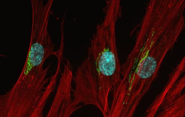
Rabbit IgG (H+L) Highly Cross-Adsorbed Secondary Antibody (A32731) in IF
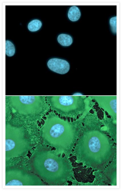
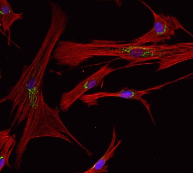
Immunofluorescent analysis of Grasp65 in HeLa cells. The cells were fixed with 4% formaldehyde for 15 mins, permeabilized with 0.25% Triton X-100 in PBS for 10 mins, and blocked with 3% BSA in PBS for 30 mins at RT. Cells were stained with a Grasp65 rabbit polyclonal antibody (Product # PA3-910) at a dilution of 1:200 in 3% BSA in PBS for 1 hr at RT, and then incubated with Invitrogen Alexa Fluor Plus 488 goat anti-rabbit IgG secondary antibody (Product # A32723) at a dilution of 1:1000 for 1 hr at RT. Nuclei were stained with Hoechst 33342 (product # H3570). The image contains overlay of Grasp65 (green) and nuclei (blue). Images were taken on a Zeiss LSM 710 confocal microscope at 40X magnification.

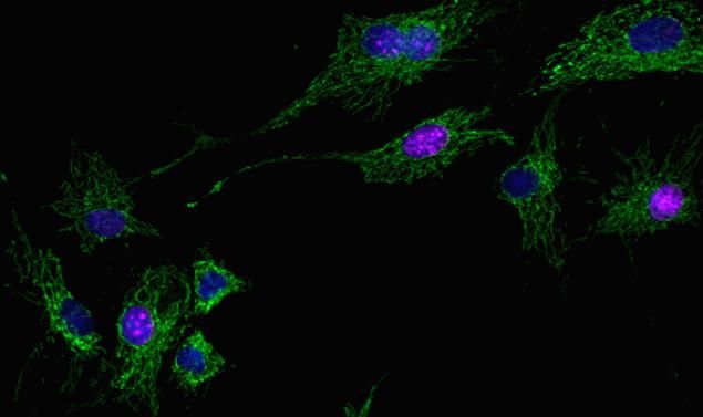
Rabbit IgG (H+L) Highly Cross-Adsorbed Secondary Antibody (A32733) in IF
Immunofluorescent analysis of postsynaptic density protein (PSD-95) in E18 Sparague Dawley primary cortical neuronal cells. The cells were fixed with 4% formaldehyde for 15 mins, permeabilized with 0.25% Triton X-100 in PBS for 10 mins, and blocked with 3% BSA in PBS for 30 mins at RT. Cells were stained with a PSD-95 rabbit polyclonal antibody (Product # 51-6900) at a dilution of 1:200 in 3% BSA in PBS for 1 hr at RT, and then incubated with Invitrogen Alexa Fluor Plus 647 goat anti-rabbit IgG secondary antibody (Product # A32733) at a dilution of 1:1000 for 1 hr at RT. Nuclei were stained with Hoechst 33342 (product # H3570). The image contains overlay of neuronal PSD-95 (far red) and nuclei (blue). Images were taken on a Zeiss LSM 710 confocal microscope at 40X magnification.
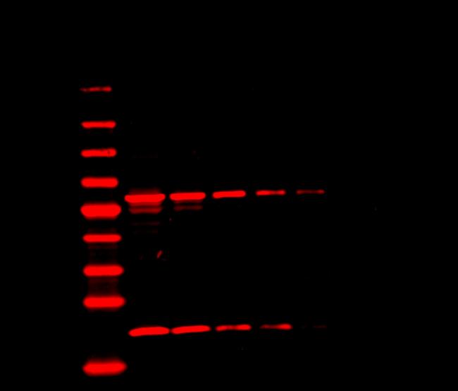
Mouse IgG (H+L) Highly Cross-Adsorbed Secondary Antibody (A32729) in WB
Western blot analysis of protein disulphide-isomerase (PDI) and progesterone receptor complex (p23) was performed by loading 3-fold serial dilutions of A431 whole cell lysate (starting at 15ug) and 2ul of the PageRuler Prestained NIR Protein Ladder (Product # 26635) per well onto a 4-20% Tris-HCl polyacrylamide gel. Proteins were transferred to Nitrocellulose Membranes (Product # 88018) and blocked with Fluorescence Blocker for 30 min. Membranes were probed with a PDI monoclonal antibody (Product # MA3-019) at a dilution of 1:5000 and a p23 monoclonal antibody at a dilution of 1:1000 (Product # MA3-414) overnight at 4°C on a rocking platform, washed with TBST, and probed with an Invitrogen Alexa Fluor Plus 680 Goat anti-Mouse IgG secondary antibody (Product # A32729) at dilutions of 1:40,000 for 45 minutes. Blots were imaged on an Infrared fluorescence imaging system.

Rabbit IgG (H+L) Cross-Adsorbed Secondary Antibody (A-11008) in IF
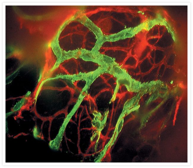
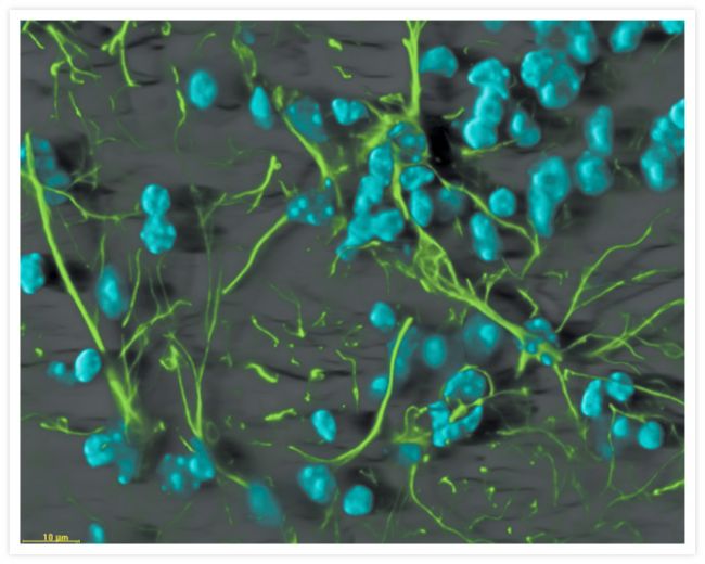
Mice were transcardially perfused with phosphate-buffered saline followed by 4% paraformaldehyde in phosphate buffer. Thirty µm serial sections were cut in a freezing microtome and transferred to phosphate-buffered saline. Free-floating sections were incubated with 1% hydrogen peroxide to quench endogenous peroxidase activity, blocked in 5% normal goat serum, then stained with a rabbit polyclonal antibody to calbindin D-28K (Chemicon) at a 1:1000 dilution. After washing, sections were incubated with Alexa Fluor® 488 goat anti-rabbit IgG antibody (Prod # A11008) at 5 µg/mL (upper panel) or HRP-goat anti-rabbit IgG antibody at 1 µg/mL, followed by Alexa Fluor® 488 tyramide (in TSA Kit #12, T20922; lower panel). Sections were washed, mounted on slides, coverslipped with ProLong® antifade reagent (in Kit P7481) and imaged under identical conditions (10X magnification, 250 millisecond exposure) using a bandpass filter set appropriate for fluorescein (FITC).

Rabbit IgG (H+L) Highly Cross-Adsorbed Secondary Antibody (A32734) in WB
Western blot analysis of Heat Shock Protein 90 (Hsp90) and histone deacetylase 1 (HDAC1) was performed by loading 3-fold serial dilutions of A431 whole cell lysate (starting at 15ug) and 2ul of the PageRuler Prestained NIR Protein Ladder (Product # 26635) per well onto a 4-20% Tris-HCl polyacrylamide gel. Proteins were transferred to Nitrocellulose Membranes (Product # 88018) and blocked with Fluorescence Blocker for 30 min. Membranes were probed with a Hsp90 polyclonal antibody (Product # PA3-013) at a dilution of 1:5000 and an HDAC1 polyclonal antibody at a dilution of 1:5000 (Product # PA1-1110) overnight at 4°C on a rocking platform, washed with TBST, and probed with an Invitrogen Alexa Fluor Plus 680 Goat anti-Rabbit IgG secondary antibody (Product # A32734) at dilutions of 1:40,000 for 45 minutes. Blots were imaged on an Infrared fluorescence imaging system.

Mouse IgG (H+L) Highly Cross-Adsorbed Secondary Antibody (A32730) in WB
Western blot analysis of protein disulphide-isomerase (PDI) and progesterone receptor complex (p23) was performed by loading 3-fold serial dilutions of A431 whole cell lysate (starting at 15ug) and 2ul of the PageRuler Prestained NIR Protein Ladder (Product # 26635) per well onto a 4-20% Tris-HCl polyacrylamide gel. Proteins were transferred to Nitrocellulose Membranes (Product # 88018) and blocked with Fluorescence Blocker for 30 min. Membranes were probed with a PDI monoclonal antibody (Product # MA3-019) at a dilution of 1:5000 and a p23 monoclonal antibody at a dilution of 1:1000 (Product # MA3-414) overnight at 4°C on a rocking platform, washed with TBST, and probed with an Invitrogen Alexa Fluor Plus 800 Goat anti-Mouse IgG secondary antibody (Product # A32730) at dilutions of 1:40,000 for 45 minutes. Blots were imaged on an Infrared fluorescence imaging system.

Rabbit IgG (H+L) Highly Cross-Adsorbed Secondary Antibody (A32735) in WB
Western blot analysis of Heat Shock Protein 90 (Hsp90) and histone deacetylase 1 (HDAC1) was performed by loading 3-fold serial dilutions of A431 whole cell lysate (starting at 15ug) and 2ul of the PageRuler Prestained NIR Protein Ladder (Product # 26635) per well onto a 4-20% Tris-HCl polyacrylamide gel. Proteins were transferred to Nitrocellulose Membranes (Product # 88018) and blocked with Fluorescence Blocker for 30 min. Membranes were probed with a Hsp90 polyclonal antibody (Product # PA3-013) at a dilution of 1:5000 and an HDAC1 polyclonal antibody at a dilution of 1:5000 (Product # PA1-1110) overnight at 4°C on a rocking platform, washed with TBST, and probed with an Invitrogen Alexa Fluor Plus 800 Goat anti-Rabbit IgG secondary antibody (Product # A32735) at dilutions of 1:40,000 for 45 minutes. Blots were imaged on an Infrared fluorescence imaging system.

Mouse IgG (H+L) Highly Cross-Adsorbed Secondary Antibody (A-11029) in IF
Simultaneous detection of expression of five genes in a whole-mount Drosophila embryo by fluorescence in situ hybridization (FISH) with five RNA probes. Red: sog labeled using aminoallyl UTP (Prod # A21663, A32765) and Alexa Fluor® 647 succinimidyl ester (Prod # A20006, A20106). Green: ind labeled with DNP, followed by rabbit anti-dinitrophenyl-KLH IgG antibody (Prod # A6430) prelabeled with the Zenon® Alexa Fluor® 555 Rabbit IgG Labeling Kit (Prod # Z25305). Blue: en labeled with biotin and detected with HRP-streptavidin and Alexa Fluor® 405 tyramide (TSA™ Kit #39, T30952). Yellow: wg labeled with digoxigenin and detected with sheep anti-digoxigenin IgG antibody and Alexa Fluor® 594 Donkey Anti-Sheep IgG antibody (Prod # A11016). Magenta: msh labeled with fluorescein and detected with mouse anti-fluorescein/Oregon Green® IgG<sub>2a</sub> antibody (Prod # A6421) and Alexa Fluor® 488 Goat Anti-Mouse IgG antibody (Prod # A11001, A11029). Image contributed by Dave Kosman and Ethan Bier, University of California, San Diego.

Mouse IgG (H+L) Highly Cross-Adsorbed Secondary Antibody (A-11032) in IF
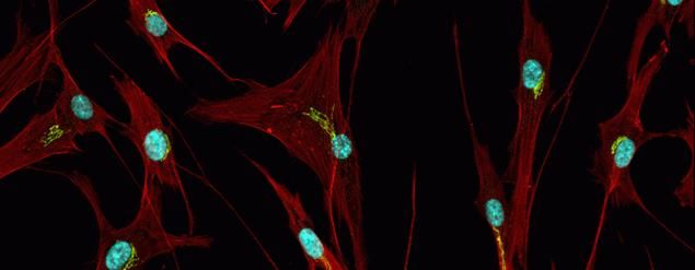
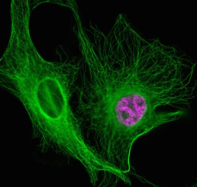
Immunofluorescence was performed on methanol fixed HeLa cells for detection of BNIP3 using Anti-BNIP3 (8HCLC) Recombinant Rabbit Oligoclonal Antibody (Product# 710728, 2ug/ml), alpha-Tubulin Monoclonal Antibody (Product# 322500, 1ug/ml) and labeled with Goat anti-Rabbit IgG (H+L) Superclonal™ Secondary Antibody, Alexa Fluor® 488 conjugate (Product # A27034, 1:2000), Goat anti-Mouse IgG Secondary Antibody, Alexa Fluor® 594 conjugate (Product # A11032, 1:400) respectively. Panel a) shows representative cells that were stained for detection and localization of BNIP3 protein (green), Panel b) is stained for nuclei (blue) using SlowFade® Gold Antifade Mountant with DAPI (Product # S36938,). Panel c) represents cytoskeletal alpha-tubulin staining (red). Panel d) is a composite image of Panels a, b and c clearly demonstrating cytoplasmic localization of BNIP3. Panel e) represents merged image of untreated cells with no signal Panel f) represents control cells with no primary Antibody to assess background.

Mouse IgG (H+L) Highly Cross-Adsorbed Secondary Antibody (A-21424) in IF
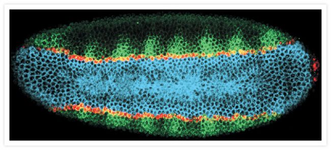
Four-color fluorescence in situ hybridization on a Drosophila embryo. A late blastoderm stage (nuclear cycle 14) embryo was probed with four different RNA probes. Blue: sog labeled with DNP, followed by a rabbit anti-dinitrophenyl-KLH IgG antibody (Prod # A6430) detected with an Alexa Fluor® 647 chicken anti-rabbit IgG antibody (Prod # A21443). Green: ind labeled with biotin, followed by streptavidin HRP and Alexa Fluor® 350 tyramide (TSA Kit #27, Prod # T20937). Red: msh labeled with digoxigenin followed by sheep anti-digoxigenin antibody detected with an Alexa Fluor® 488 donkey anti-sheep IgG antibody (Prod # A11015). Yellow: sna labeled with fluorescein followed by mouse anti-fluorescein antibody detected with an Alexa Fluor® 555 goat anti-mouse IgG antibody (Prod # A21424). Image contributed by Dave Kosman and Ethan Bier, University of California, San Diego.


