GATA4 Polyclonal Antibody for Western Blot, IF, ICC, IHC (P) and Flow
日本天野酶试剂官网
日本Amano酶中国官网代理商


GATA4 Antibody (PA1-102) in IF
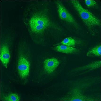
Immunofluorescent analysis of GATA4 (green) in embryoid body endoderm generated from Gibco ® Human Episomal iPSC Line grown on Geltrex® in Essential 8TM Medium. After 2 weeks in culture, EB were dissociated with TrypLETM and re-plated onto Geltrex®-coated multi-well plates. Cells were fixed, permeabilized and blocked for immunostaining. Cells were stained with a GATA4 polyclonal antibody (Product # PA1-102) at a dilution of 1:100 in 3% BSA/PBS blocking buffer overnight at 4°C, and then incubated with Alexa Fluor® 488 donkey anti rabbit antibody (Product # A21206) at a 1:500 dilution in conjunction with NucBlue® Fixed Cell Ready Probes® Reagent. After another 3 washes, images were taken on EVOS®Floid® Cell Imaging system at 10X magnification.

alpha Tubulin Antibody (A11126) in IF
Immunofluorescence analysis of Alpha-Tubulin was done on 70% confluent log phase HeLa cells. The cells were fixed with 4% paraformaldehyde for 15 minutes, permeabilized with 0.25% Triton™ X-100 for 10 minutes, and blocked with 5% BSA for 1 hour at room temperature. The cells were labeled with Alpha-Tubulin Mouse monoclonal Antibody (A11126) at 1µg/mL in 1% BSA and incubated for 3 hours at room temperature and then labeled with Alexa Flour 488 Rabbit Anti-Mouse IgG Secondary Antibody (A11059) at a dilution of 1:400 for 30 minutes at room temperature (Panel a: green). Nuclei (Panel b: blue) were stained with SlowFade® Gold Antifade Mountant DAPI (S36938). F-actin (Panel c: red) was stained with Alexa Fluor 594 Phalloidin (A12381). Panel d is a merged image showing cytoplasmic localization. Panel e shows no primary antibody control. The images were captured at 20X magnification.

LIN28A Antibody (PA1-096) in IF
Immunofluorescent analysis of LIN28 (green) in Hela cells. Formalin-fixed cells were permeabilized with 0.1% Triton X-100 in TBS for 5-10 minutes at room temperature and blocked with 3% BSA-PBS for 30 minutes at room temperature. Cells were probed with a LIN28 polyclonal antibody (Product # PA1-096 ) at a dilution of 1:100 and incubated overnight in a humidified chamber. Cells were washed with PBST and incubated with a DyLight-conjugated secondary antibody for 45 minutes at room temperature in the dark. F-actin (red) was stained with a fluorescent phalloidin and nuclei (blue) were stained with DAPI. Images were taken at a 60X magnification.

PDI Antibody (MA3-019) in IF
Immunofluorescent analysis of Phalloidin (orange) and PDI (green) in NIH 3T3 cells. Formalin fixed cells were permeabilized with 0.1% Triton X-100 in PBS for 10 minutes at room temperature and blocked with 2% BSA (Product # 37525) in PBS + 0.1% Triton X-100 for 30 minutes at room temperature. Cells were probed with a PDI monoclonal antibody (Product # MA3-019) at a dilution of 1:75 for at least 1 hour at room temperature, washed with PBS, and incubated with DyLight 488 goat anti-mouse IgG secondary antibody (Product # 35502) at a dilution of 1:250 for 30 minutes at room temperature. Actin was stained with DyLight 550 Phalloidin (Product # 21835) at a dilution of 1:120 (2.5 units/ml final concentration) and nuclei (blue) were stained with Hoechst (Product # 62249) at a concentration of 1ug/ml for 30 minutes. Images were taken on a Zeiss Axio Observer Z1 microscope at 20X magnification.

Calreticulin Antibody (PA3-900) in IF
Immunofluorescent analysis of Calreticulin (green) U2OS cells. Formalin fixed cells were permeabilized with 0.1% Triton X-100 in PBS for 10 minutes at room temperature and blocked with 2% BSA in PBS 0.1% triton-X (Product #37525) for 30 minutes at room temperature. Cells were probed with a rabbit polyclonal antibody recognizing Calreticulin (Product # PA3-900) at a dilution of 1:50 for at least 1 hour at room temperature. Cells were washed with PBS and incubated with DyLight 488 goat-anti-rabbit IgG secondary antibody (Product #35552) at a dilution of 1:250 for 30 minutes at room temperature. Actin filaments (red) were stained with DyLight 554-Phalloidin (Product# 21834) at a dilution of 1:300 (i.e. 1unit/mL final concentration) in PBS and incubated for 30 min including. Nuclei (blue) were stained with Hoechst 33342 dye (Product# 62249) at a dilution of 1µg/mL. Images were taken on a Thermo Scientific ArrayScan VTI at 20X magnification.

beta Tubulin Loading Control Antibody (MA5-16308) in IF
Immunofluorescent analysis of Beta-Tubulin (red) in HEK293T cells. Cells fixed in 4% formaldehyde were permeabilized and blocked with 1X PBS containing 5% BSA and 0.3% Triton X-100 for 1 hour at room temperature. Cells were probed with a Beta-Tubulin monoclonal antibody (Product # MA5-16308) at a dilution of 1:100 overnight at 4°C in 1X PBS containing 1% BSA and 0.3% Triton X-100, washed with 1X PBS, and incubated with a fluorophore-conjugated goat anti-mouse IgG secondary antibody at a dilution of 1:200 for 1 hour at room temperature. Nuclei (blue) were stained with DAPI. Images were taken on a Leica DM1000 microscope at 40X magnification. Data courtesy of the Innovators Program.

SOX2 Antibody (PA1-094) in IF
Immunofluorescent analysis of SOX2 using anti-SOX2 polyclonal antibody (Product# PA1-094) shows specific expression in human embryonal carcinoma NTERA-2 cells (shown in green) but not in negative control HeLa cells. Formalin fixed cells were permeabilized with 0.1% Triton X-100 in TBS for 10 minutes at room temperature. Cells were blocked with 1% Blocker BSA (Product #37525) for 15 minutes at room temperature. Cells were probed with a rabbit polyclonal antibody recognizing SOX2 (Product# PA1-094), at a dilution of 1:200 for at least 1 hour at room temperature. Cells were washed with PBS and incubated with DyLight 488 goat-anti-rabbit IgG secondary antibody (Product# 35552) at a dilution of 1:400 for 30 minutes at room temperature. F-Actin (red) was stained with DY-547 phalloidin, nuclei (blue) were stained with Hoechst 33342 dye (Product# 62249). Images were taken on a Thermo Scientific ArrayScan at 20X magnification.

GAPDH Loading Control Antibody (MA5-15738) in IF
Immunofluorescent analysis of GAPDH (green) in HeLa. Formalin-fixed cells were permeabilized with 0.1% Triton X-100 in TBS for 10 minutes at room temperature and blocked with 5% normal goat serum (Product # 31873) for 15 minutes at room temperature. Cells were probed with a GAPDH monoclonal antibody (Product # MA5-15738) at a dilution of 1:100 for at least 1 hour at room temperature, washed with PBS, and incubated with DyLight 488 goat anti-mouse IgG secondary antibody (Product # 35502) at a dilution of 1:400 for 30 minutes at room temperature. Nuclei (blue) were stained with Hoechst 33342 dye (Product # 62249). Images were taken on a Thermo Scientific ArrayScan or ToxInsight Instrument at 20X magnification.

LIN28A Antibody (PA1-096X) in IF
Immunofluorescent analysis of Lin28 (red) in H9 embryonic stem cells grown for a few days on Matrigel-coated chamber slides. Cells fixed in 4% paraformaldehyde were permeabilized with 0.1% Triton X-100 for 15 minutes at room temperature. Cells were probed with a Lin28 polyclonal antibody (Product # PA1-096) at a dilution of 1:200 overnight at 4°C, washed with PBST, and incubated with a fluorescently-conjugated secondary antibody at a dilution of 1:100 for 1 hour at room temperature. Nuclei (blue) were stained with DAPI and cells were analyzed by fluorescence microscopy at 20X magnification.

SOX2 Antibody (MA1-014) in IF
Immunofluorescent analysis of Sox2 (green) in H9 embryonic stem cells grown for a few days on Matrigel-coated chamber slides. Cells fixed in 4% paraformaldehyde were permeabilized with 0.1% Triton X-100 for 15 minutes at room temperature. Cells were probed with a Sox2 monoclonal antibody (Product # MA1-014) at a dilution of 1:200 overnight at 4°C, washed with PBST, and incubated with a fluorescein-conjugated secondary antibody at a dilution of 1:100 for 1 hour at room temperature. Nuclei (blue) were stained with DAPI and cells were analyzed by fluorescence microscopy at 20X magnification.


