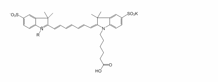
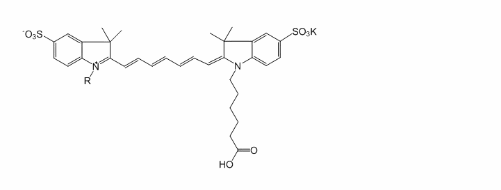
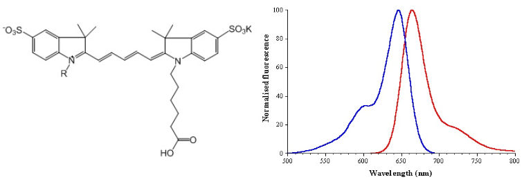
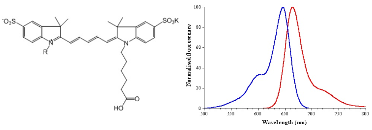
Cy3 SE
基本信息
| 产品名称 | Cy3 SE |
|---|---|
| 英文名称 | Cy3 SE |
| 运输条件 | 超低温冰袋运输 |
一般描述
产品简介
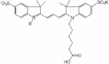
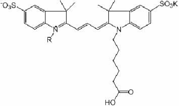
菁染料是性能优良的荧光标记染料,摩尔吸光系数在荧光染料中是最高的,其琥珀酰亚胺酯是最常用的脂肪氨基标记试剂,广泛用于蛋白、抗体、核酸及其他生物分子的标记和检测。通过改变次甲基链的长度, 可改变其荧光发射波长,每增加一个双键,按照Huoffman规则正好红移约100nm。
菁染料Cy3和Cy5已成为基因芯片的首选荧光标记物;另外,Cy5, Cy5.5和Cy7的吸收在近红外区背景非常低,是荧光强度最高、最稳定的长波长染料。特别适合于活体小动物体内成像代替放射性元素。
产品参数
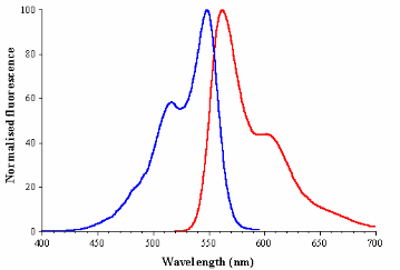
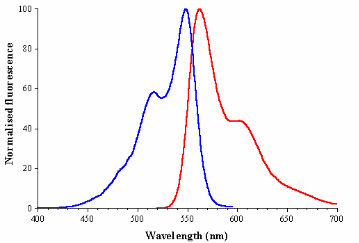
Ex(nm) 548 Em(nm) 562 Solvent: Water or DMSO
菁染料琥珀酰亚胺酯(CyDye NHS)的标记
菁染料和生物分子的比例F/P=4~12之间荧光强度最高,F/P值过高荧光探针会自我淬灭并影响生物分子的生物活性,标记生物分子最好是用单琥珀酰亚胺酯,但是用双修饰的CyDye NHS并没有发现交联。CyDye NHS标记抗体在pH (8.5~9.4)时10分钟F/P可达5~6,而在pH 7.0几乎不反应。我们用不同比例的Cy3标记anti-glutathione-S-transferase (GST)多克隆抗体发现用1:1, 5:1, 10:1 和20:1标记时得到的F/P值分别是0.28:1, 1.16:1, 2.3:1 和4.6:1。
操作步骤
水溶性Cy3 NHS标记anti-GST多克隆抗体
市场上买到的抗体如果含有其它蛋白(例如serum albumin或gelatin等)或带氨基的缓冲液会影响标记,在标记前需纯化。
1. 在1 L 0.15 M NaCl 溶液中透析anti-GST抗体(浓度为0.5 mL at 3 mg/mL),常温透析4小时。
2. 在4°C用新鲜的1 L 0.15 M NaCl 溶液再透析过夜。
3. 第二天用1 L 0.1 M NaHCO3(pH 8.3)透析4小时。
4. 用0.22 μm注射器过滤头过滤抗体溶液。
5. 用0.1 M NaHCO3稀释少量的抗体,在280nm处测其紫外吸收值计算标记抗体的总量(IgG antibody摩尔吸光系数170 000 M-1cm-1at 280 nm).
6. 用DMSO配置Cy3 NHS (MW 765.95)溶液浓度为10 mg/mL;计算所需体积以得到想要的CyDye NHS 和抗体的比值(例如20:1),然后慢慢将其加入到抗体溶液中,同时在暗处常温缓慢搅拌45分钟。
7. 用1 L 0.15 M NaCl 溶液常温避光透析4小时除去未标记上的Cy3。
8. 在4°C用新鲜的1 L 0.15 M NaCl 溶液再避光透析过夜。
9. 用1 L of 0.01 M PBS/ 0.01% 叠氮化钠溶液常温避光透析4小时, 在4°C常温避光再次透析过夜。
10. 用0.22 μm注射器过滤头过滤抗体溶液。
11. 用0.01 M PBS/0.01 % 叠氮化钠整数倍稀释标记抗体溶液,测量280nm(蛋白)和552nm(Cy)处的紫外可见吸光度。
12. 产品冷冻干燥成粉末或在0.01 M PBS/ 0.01% 叠氮化钠溶液中,-20℃避光储存。
F/P计算:
Cy3在552nm摩尔吸光系数为150000 M-1cm-1;此蛋白在280nm的摩尔吸光系数为170 000 M-1cm-1;不同蛋白的摩尔吸光系数不一样;.Cy3 染料本身在280 nm 的吸收是552nm处的8%。按以下公式计算F/P值。
[Cy3] = A552/150000
[antibody] = {A280- (0.08×A552)}/170000
F/P final= [Cy3]/[antibody]= {1.13×A552}/ {A280- (0.08×A552)}
Product Introduction
Cyanine dye is a fluorescent labeling dye with excellent performance. Its molar absorption coefficient is the highest among fluorescent dyes. Its succinimidyl ester is the most commonly used aliphatic amino group labeling reagent and is widely used for labeling proteins, antibodies, nucleic acids and other biological molecules. And detection. By changing the length of the methine chain, the fluorescence emission wavelength can be changed. For each double bond added, the red shift is exactly 100 nm according to the Huoffman rule.
The cyanine dyes Cy3 and Cy5 have become the first choice fluorescent markers for gene chips; in addition, the absorption of Cy5, Cy5.5 and Cy7 is very low in the near-infrared region, and they are the long-wavelength dyes with the highest fluorescence intensity and the most stable. It is especially suitable for in vivo imaging of small living animals instead of radioactive elements.
Product parameter
Ex(nm) 548 Em(nm) 562 Solvent: Water or DMSO
Labeling of cyanine dye succinimide ester (CyDye NHS)
The ratio of cyanine dye and biomolecule is between F/P=4~12. The fluorescence intensity is the highest. If the F/P value is too high, the fluorescent probe will self-quench and affect the biological activity of biomolecules. It is best to use single amber to label biomolecules. Imide ester, but CyDye NHS modified with double did not find cross-linking. CyDye NHS-labeled antibody can reach 5-6 F/P in 10 minutes at pH (8.5-9.4), but hardly reacts at pH 7.0. We used different ratios of Cy3 to label anti-glutathione-S-transferase (GST) polyclonal antibodies and found that the F/P values obtained when labeled with 1:1, 5:1, 10:1 and 20:1 were 0.28:1 , 1.16:1, 2.3:1 and 4.6:1.
Steps
Water-soluble Cy3 NHS labeled anti-GST polyclonal antibody
Antibodies bought on the market containing other proteins (such as serum albumin or gelatin, etc.) or buffers with amino groups will affect the labeling, and need to be purified before labeling.
1. Dialyze the anti-GST antibody (concentration 0.5 mL at 3 mg/mL) in 1 L 0.15 M NaCl solution for 4 hours at room temperature.
2. Dialysis with fresh 1 L 0.15 M NaCl solution at 4°C overnight.
3. Dialysis with 1 L 0.1 M NaHCO3 (pH 8.3) for 4 hours on the second day.
4. Filter the antibody solution with a 0.22 μm syringe filter.
5. Dilute a small amount of antibody with 0.1 M NaHCO3, measure its UV absorption value at 280nm to calculate the total amount of labeled antibody (IgG antibody molar absorption coefficient 170 000 M-1cm-1at 280 nm).
6. Use DMSO to configure the Cy3 NHS (MW 765.95) solution concentration to 10 mg/mL; calculate the required volume to get the desired ratio of CyDye NHS to antibody (for example, 20:1), and then slowly add it to the antibody solution In the meantime, stir slowly for 45 minutes at room temperature in the dark.
7. Dialysis with 1 L 0.15 M NaCl solution at room temperature and avoid light for 4 hours to remove unlabeled Cy3.
8. Use fresh 1 L 0.15 M NaCl solution at 4°C to dialyze overnight in the dark.
9. Use 1 L of 0.01 M PBS/ 0.01% sodium azide solution to dialyze at room temperature and avoid light for 4 hours, and then dialyze again overnight at 4°C and avoid light.
10. Filter the antibody solution with a 0.22 μm syringe filter.
11. Dilute the labeled antibody solution with 0.01 M PBS/0.01% sodium azide integer times, and measure the UV-visible absorbance at 280nm (protein) and 552nm (Cy).
12. The product is freeze-dried into powder or stored in 0.01 M PBS/ 0.01% sodium azide solution at -20°C and protected from light.
F/P calculation:
The molar absorption coefficient of Cy3 at 552nm is 150000 M-1cm-1; the molar absorption coefficient of this protein at 280nm is 170 000 M-1cm-1; the molar absorption coefficients of different proteins are different; the absorption of Cy3 dye itself at 280 nm is 8% at 552nm. Calculate the F/P value according to the following formula.
[Cy3] = A552/150000
[antibody] = {A280- (0.08×A552)}/170000
F/P final= [Cy3]/[antibody]= {1.13×A552}/ {A280- (0.08×A552)}
相关属性
| 储存温度 | 避光,-20°C储存,干燥 |
|---|---|
| 品牌 | Jinpan |


